This was in the era when surgery and bacteriology were beginning to converge. Surgeons were learning that microorganisms introduced into wounds could cause infection. They were starting to ask which organisms caused the pus, necrosis and deformity they encountered in surgical disease, and whether different microorganisms could produce different patterns of disease.
German-speaking surgeons were adopting Robert Koch’s methods of bacterial culture and experimental inoculation, and some surgical departments established bacteriology laboratories of their own (Schlich, 2012). Wound infection and osteomyelitis could increasingly be investigated by isolating organisms from affected tissue and testing them experimentally (Schlich, 2012).
A counterintuitive response to infection
In his 1893 paper, Garré described a bone response that still feels counterintuitive. He described osteomyelitis with marked thickening and sclerosis of the affected bone (Garré, 1893). This was two years before Röntgen described X-rays in 1895, so Garré was describing these bone changes without radiographic imaging (Garré, 1893; Röntgen, 1895).
But in 1893 Garré had already noticed the biological oddity that is still difficult to explain: infected bone could respond by making more bone (Garré, 1893).
And then Garré inoculated himself
In the 1880s, while investigating the organisms responsible for suppurative disease, Garré inoculated the skin of his own arm with staphylococci cultured from osteomyelitis cases (Garré, 1885; Schlich, 2012).
Garré was working in Basel, where surgeon August Socin had established a bacteriology laboratory and placed Garré in charge of it. He later qualified to teach both surgery and bacteriology. Using Koch’s pure culture and inoculation methods, he investigated staphylococci isolated from osteomyelitis cases in animals and in himself. The self inoculation produced a severe local infection that became difficult to control (Garré, 1885; Schlich, 2012).
A self-inoculation experiment of this kind would be unacceptable under current research ethics. The experimental logic, however, is completely recognisable: isolate the organism, introduce it deliberately, and see whether disease follows.
The result also complicated the emerging idea that one bacterial species corresponded to one specific disease. The staphylococcus Garré had isolated from osteomyelitis cases could also produce other purulent infections, including furunculosis and panaritium. Garré concluded that the same organism could produce different forms of suppurative disease (Garré, 1885; Schlich, 2012).
Let’s have a closer look
Histological sections from experimental Staphylococcus aureus osteomyelitis show the extent of this proliferative response.
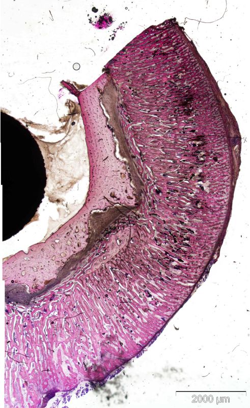
Histological overview of reactive bone formation
Histological cross section of an infected rabbit tibia showing the original cortex and extensive periosteal new bone formation. The black circular profile corresponds to the intramedullary implant. A portion of the anterior tibia had been removed for bacterial culture before histological processing.
The original cortex remains readily identifiable as the more compact, lightly stained bone surrounding the medullary cavity. Much of the original cortical structure is preserved, but its outer contour is irregular and focally interrupted by areas of cortical resorption.
Beyond this residual cortex, extensive newly formed bone extends along the periosteal surface. Compared with the original cortex, the newly formed bone has a much more open and irregular architecture and extends well beyond the original cortical boundary.
Simultaneous bone destruction and bone formation
In a rabbit tibia model of S. aureus infection, fluorochrome labelling showed excessive subperiosteal bone formation beginning during the first week and continuing throughout the four- week study period. After 28 days, infected proximal tibiae contained approximately twice the bone volume of uninfected controls, while micro-CT showed cortical lysis (Croes et al., 2017).
Cortical lysis and subperiosteal bone formation therefore occurred simultaneously in the infected tibiae (Croes et al., 2017).
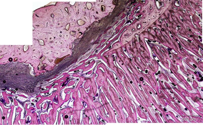
Higher magnification view of the original cortex and adjacent newly formed bone
More bone is not necessarily better bone
In experimental osteomyelitis, fracture and defect healing can remain impaired even while periosteal new bone develops elsewhere (Croes et al., 2019b).
Extensive reactive bone formation can therefore coexist with impaired structural healing and does not demonstrate restoration of normal architecture or mechanical function (Croes et al., 2019b).
Wolff’s law describes adaptation of bone to mechanical loading. In osteomyelitis, reactive periosteal bone can appear early and in locations that do not correspond to the areas requiring structural repair. In the rabbit tibia model, the adjacent ipsilateral fibula did not show the same osteogenic response as the infected tibia (Croes et al., 2017). Reactive bone can also develop away from the regions of greatest bone loss, making a simple biomechanical adaptation to osteolysis unlikely (Croes et al., 2019b).
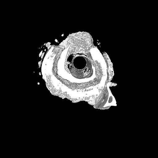
Transverse micro-CT image of an infected rabbit tibia
The residual original cortex is highlighted in yellow and the adjacent fibula in purple. The central black profile corresponds to the intramedullary implant. Extensive mineralised reactive bone extends beyond the original cortical boundary, while parts of the original cortex show irregularity and cortical loss.
Bacterial components can trigger reactive bone formation
An isolated S. aureus cell wall extract induced new bone formation and cortical thickening without the extensive cortical lysis produced by viable infection (Croes et al., 2017).
Later experiments showed that killed bacteria could also induce substantial new bone formation without reproducing the full destructive phenotype of active infection (Croes et al., 2019a). Viable bacteria were therefore not required to induce the osteogenic response. Lipoteichoic acid, a Gram-positive cell wall component and Toll-like receptor 2 agonist, was subsequently identified as one osteostimulatory bacterial stimulus (Croes et al., 2019a).
The bacterial phenotype influences how much reactive bone forms
Altering the S. aureus regulator sarA significantly reduced reactive new bone formation in a murine osteomyelitis model (Beenken et al., 2025). sarA modulates the expression of many S. aureus genes, and mutation of sarA increases extracellular protease production (Beenken et al., 2025).
Changing bacterial regulation therefore changed the amount of reactive bone formed, while the infecting species remained the same (Beenken et al., 2025).
The response is compartmentalised
Across experimental models, the region with the greatest bacterial burden is often spatially distinct from the region with the greatest new bone formation. Reactive bone may develop further from the infected focus or along the periosteal surface, while healing immediately around an infected defect remains poor (Croes et al., 2019b).
Intense inflammation close to the bacterial burden may inhibit osteogenesis, whereas less intense inflammatory signalling at more peripheral sites may permit or stimulate new bone formation (Croes et al., 2019b).
We understand bone loss better than reactive bone formation
In a 2024 murine model of S. aureus osteomyelitis, infection strongly increased RANKL expression in bone marrow stromal cells. Blocking RANKL with denosumab prevented osteoclast formation and completely prevented cortical bone destruction without changing bacterial burden (Campbell et al., 2024).
RANKL-mediated osteoclast formation was therefore required for cortical bone loss in this model. Reactive periosteal bone formation, however, persisted despite RANKL blockade, although the amount was reduced (Campbell et al., 2024).
Osteoclasts and osteoblasts stop working as a team
In physiological bone remodelling, osteoclastic resorption and osteoblastic bone formation are coupled. In a murine S. aureus osteomyelitis model, regions with a high bacterial burden showed abundant osteoclast activity but few osteoblasts, while osteoblasts remained present in adjacent remodelling bone (Campbell et al., 2024).
Bone resorption and bone formation were therefore spatially uncoupled around the infected focus (Campbell et al., 2024).
Osteoblasts help recruit immune cells
Osteoblasts are bone-forming cells, but they also respond directly to S. aureus. Exposure to live S. aureus stimulated osteoblasts to produce CCL20, a chemokine involved in recruitment of CCR6-positive immune cells (Meghwani et al., 2025). In infected mice, loss of CCL20 or CCR6 impaired T cell recruitment, while CCR6 deficient mice showed increased reactive bone formation and osteoclast numbers (Meghwani et al., 2025).
CCL20/CCR6 signalling therefore influences immune cell recruitment and the bone response during S. aureus osteomyelitis, although why CCR6 deficiency increases reactive bone formation remains unresolved (Meghwani et al., 2025).
Garré saw in 1893 what we are still trying to explain
Garré described the proliferative bone phenotype in 1893 (Garré, 1893). More than 130 years later, the mechanisms of bone destruction are much better understood, but what starts the periosteal bone formation Garré described is still unknown (Campbell et al., 2024).
Histological and micro-CT images adapted from the experimental work reported in Boot et al. (2020), using original image files from the author’s research archive.
References
Beenken KE, Campbell MJ, Smeltzer MS. The ability of sarA to limit protease production plays a key role in the pathogenesis of Staphylococcus aureus osteomyelitis irrespective of the functional status of agr. Infection and Immunity. 2025;93(1):e00473-24.
Boot W, Vogely HC, Nikkels PGJ, Pouran B, van Rijen MHP, Ekkelenkamp MB, Hänsch GM, Dhert WJA, Gawlitta D. Prophylaxis of implant-related infections by local release of vancomycin from a hydrogel in rabbits. European Cells & Materials. 2020;39:108–120.
Campbell MJ, Bustamante-Gomez C, Fu Q, Beenken KE, Reyes-Pardo H, Smeltzer MS, O'Brien CA. RANKL-mediated osteoclast formation is required for bone loss in a murine model of Staphylococcus aureus osteomyelitis. Bone. 2024;187:117181.
Croes M, Boot W, Kruyt MC, Weinans H, Pouran B, van der Helm YJM, Gawlitta D, Vogely HC, Alblas J, Dhert WJA, Öner FC. Inflammation-Induced Osteogenesis in a Rabbit Tibia Model. Tissue Engineering Part C: Methods. 2017;23(11):673–685.
Croes M, Kruyt MC, Boot W, Pouran B, Braham MVJ, Pakpahan SA, Weinans H, Vogely HC, Fluit AC, Dhert WJA, Alblas J, Öner FC. The role of bacterial stimuli in inflammation-driven bone formation. European Cells & Materials. 2019;37:402–419.
Croes M, van der Wal BCH, Vogely HC. Impact of Bacterial Infections on Osteogenesis: Evidence From In Vivo Studies. Journal of Orthopaedic Research. 2019;37(10):2067–2076.
Garré C. Zur Aetiologie acut eitriger Entzündungen. Fortschritte der Medicin. 1885;3:165–172.
Garré C. Ueber besondere Formen und Folgezustände der akuten infektiösen Osteomyelitis. Beiträge zur klinischen Chirurgie. 1893;10:241–298.
Meghwani H, Rangel-Moreno J, Sandercock KM, Saito M, McDonald KA, Kraft CM, Constantine R, Lenigk S, Rodriguez A, Kates SL, Jonason JH, Schwarz EM, Muthukrishnan G. CCL20/CCR6 signaling modulates disease severity during the establishment of Staphylococcus aureus osteomyelitis. mBio. 2025;16(10):e01413-25.
Röntgen WC. Ueber eine neue Art von Strahlen. Sitzungsberichte der Physikalisch-Medicinischen Gesellschaft zu Würzburg. 1895;(9):132–141.
Schlich T. Asepsis and Bacteriology: A Realignment of Surgery and Laboratory Science. Medical History. 2012;56(3):308–334.